11/07/2023 0 Comments
What to Expect during a CT or MRI Scan for Your Pet
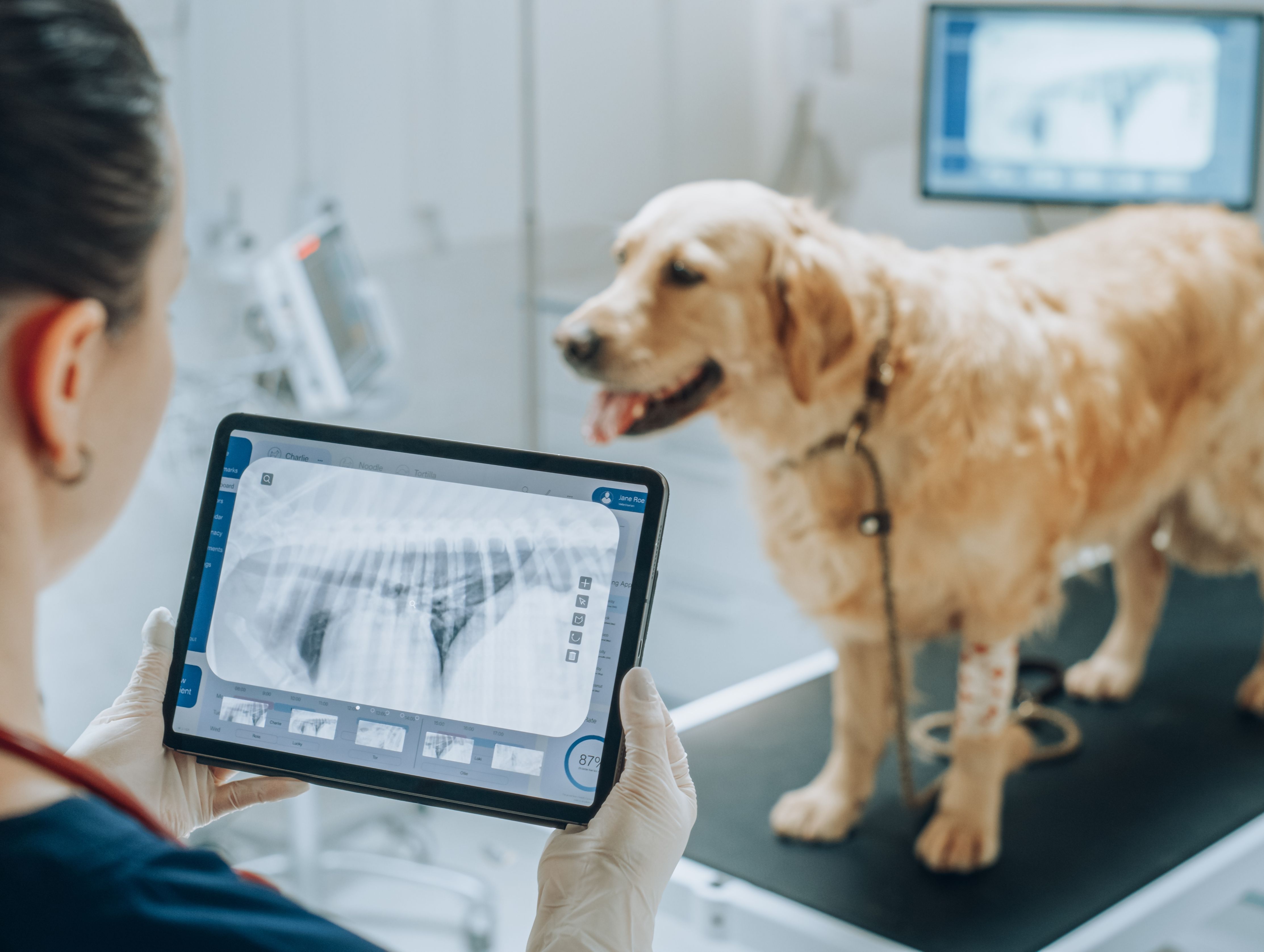
CT scans and MRI scans have revolutionised veterinary medicine. These non-invasive procedures provide detailed images of the internal structures, aiding veterinarians in making accurate diagnoses and developing appropriate treatment plans.
It was natural to be concerned and curious about the MRI scan and CT scan process, so it is helpful to know what to expect, so you can properly prepare your pet for the assessment.
What are CT and MRI Scans, and what are the differences?
CT (Computed Tomography) and MRI (Magnetic Resonance Imaging) scans are advanced imaging techniques used to visualise the internal structures of the body. While they serve a similar purpose, they differ in the technology used to create the images. CT scans use X-rays and computer processing to generate cross-sectional images, whereas MRI scans use powerful magnets and radio waves to produce detailed images. They are used in a number of veterinary medicinal areas such as oncology, cardiology and orthopaedics.
Before the scan
Before the scan, you will receive specific instructions from the veterinary clinic. These instructions will vary in accordance with what the vet is looking out for to give your pet a diagnosis. Instructions should include fasting requirements, restrictions on medication intake, and removal of any metal objects such as collars. It is important to follow these guidelines to ensure the best possible results from the scan.
MRI Sedation and Anaesthesia
In some cases, your beloved pet may require sedation or anaesthesia to keep them calm and still during the scanning process. This is particularly important for MRI scans, as the procedure can take a good amount of time. Veterinarians will assess the pet's temperament and health condition to determine if sedation or anaesthesia is necessary.
The CT Scan Procedure
During a CT scan, the pet is placed on a table that moves through a circular opening. X-ray beams are projected through the body from various angles, and detectors on the opposite side measure the amount of radiation absorbed. A computer then processes the data and generates detailed cross-sectional images, providing valuable information about the internal structures.
The MRI Scan Procedure
MRI scans require the pet to lie on a table that moves into a cylindrical machine. The machine uses powerful magnets and radio waves to create detailed images of the body's structures. The procedure is quite noisy, so to minimise stress and movement, pets are usually placed under anaesthesia, as mentioned above. Your pet's safety will be monitored closely whilst under anaesthesia throughout the procedure.
All finished and time to go home
Once the scanning procedure is complete and your brave pet has come around after the anaesthesia, the veterinarian will review the images and interpret the findings. The results will help determine the presence of any abnormalities, such as tumours, fractures, or organ dysfunction.
Comments
Leave a comment